Description
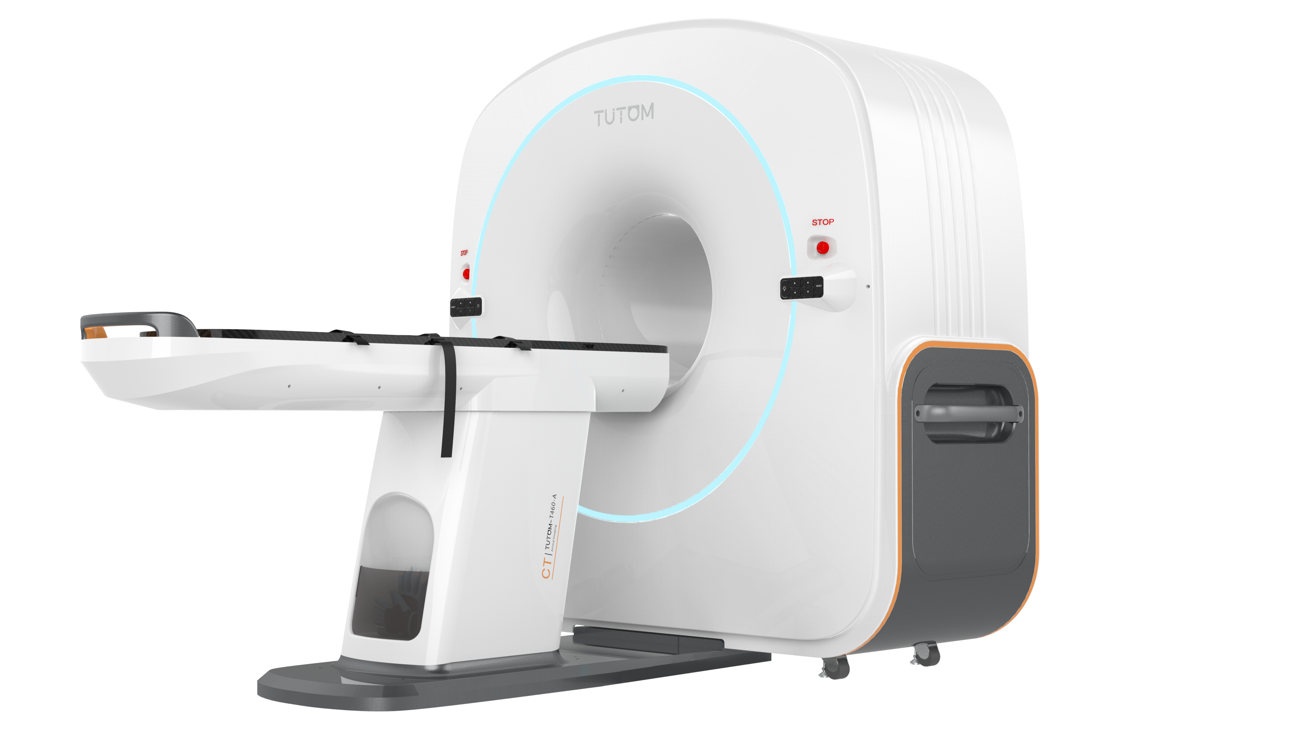
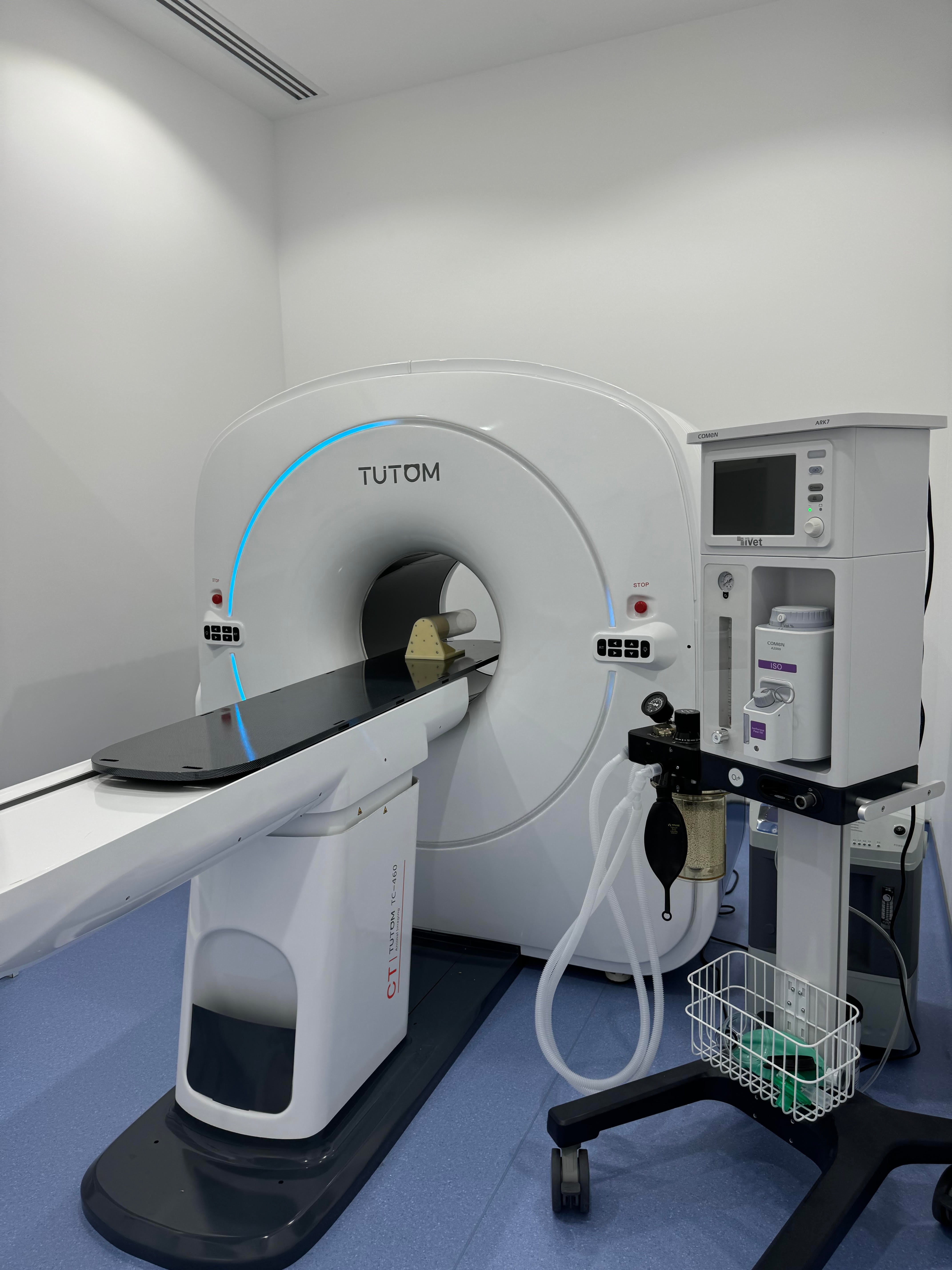
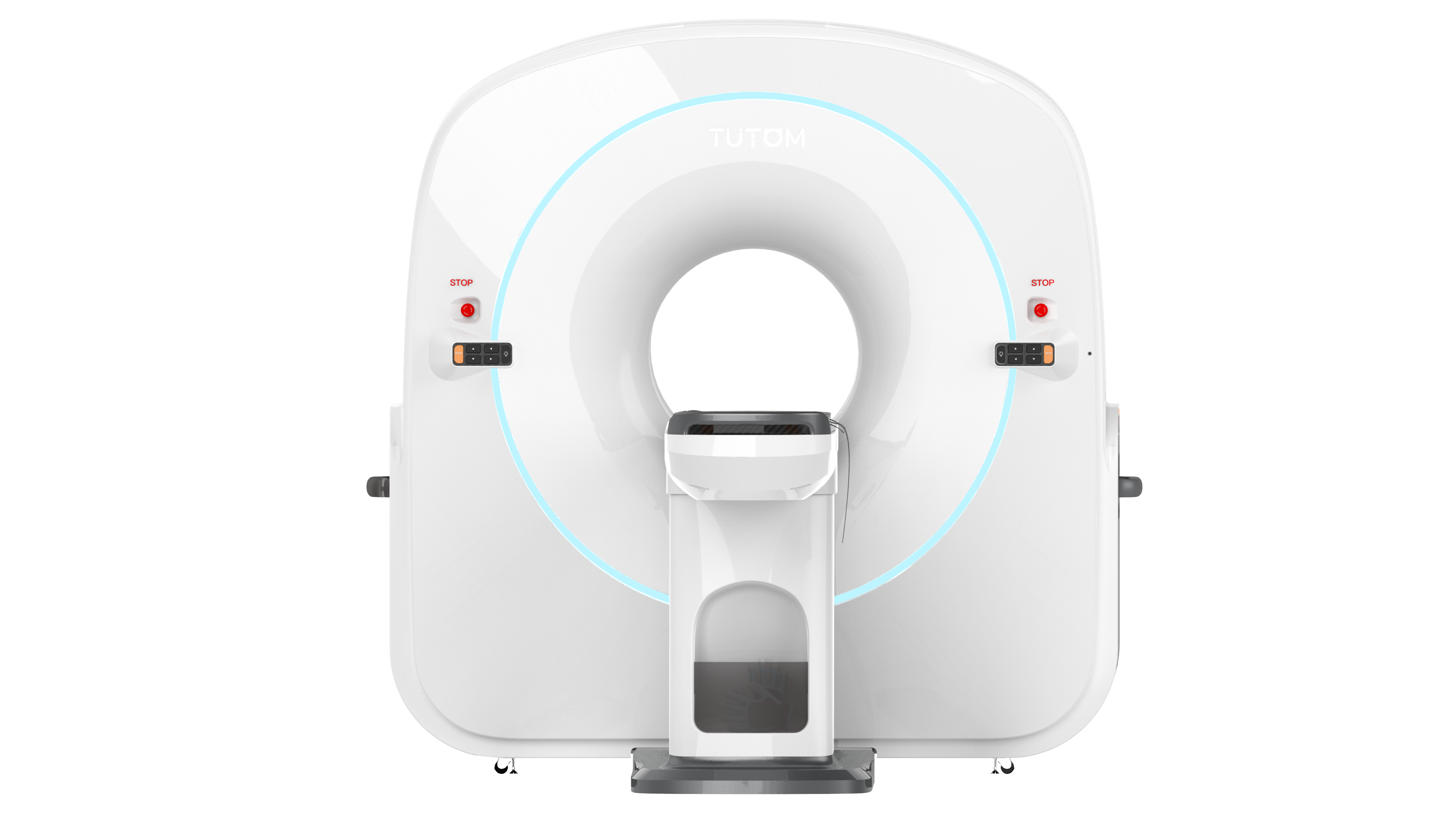
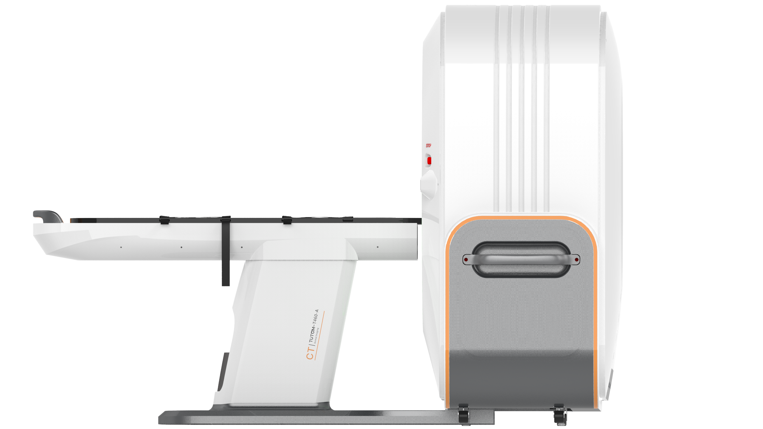
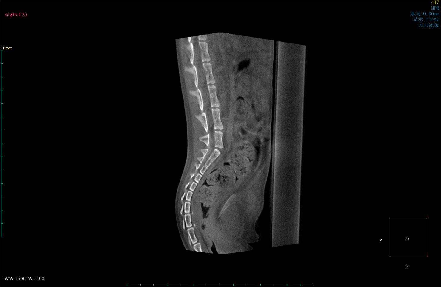
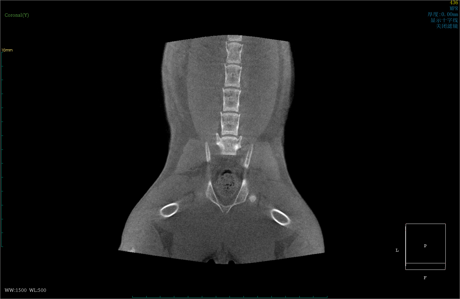
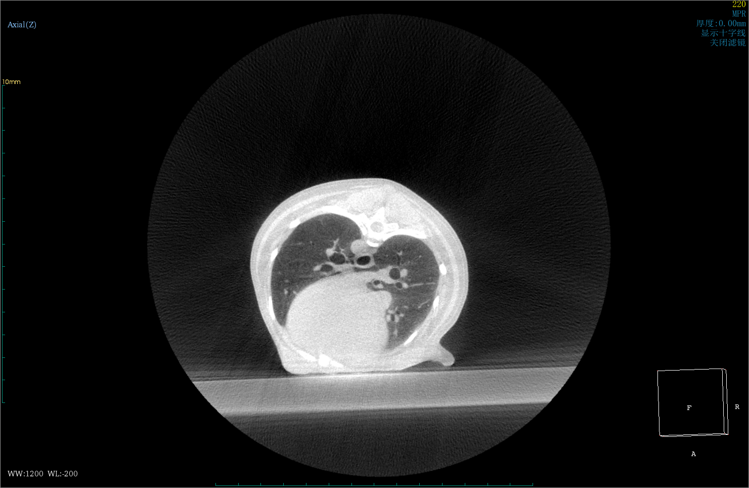
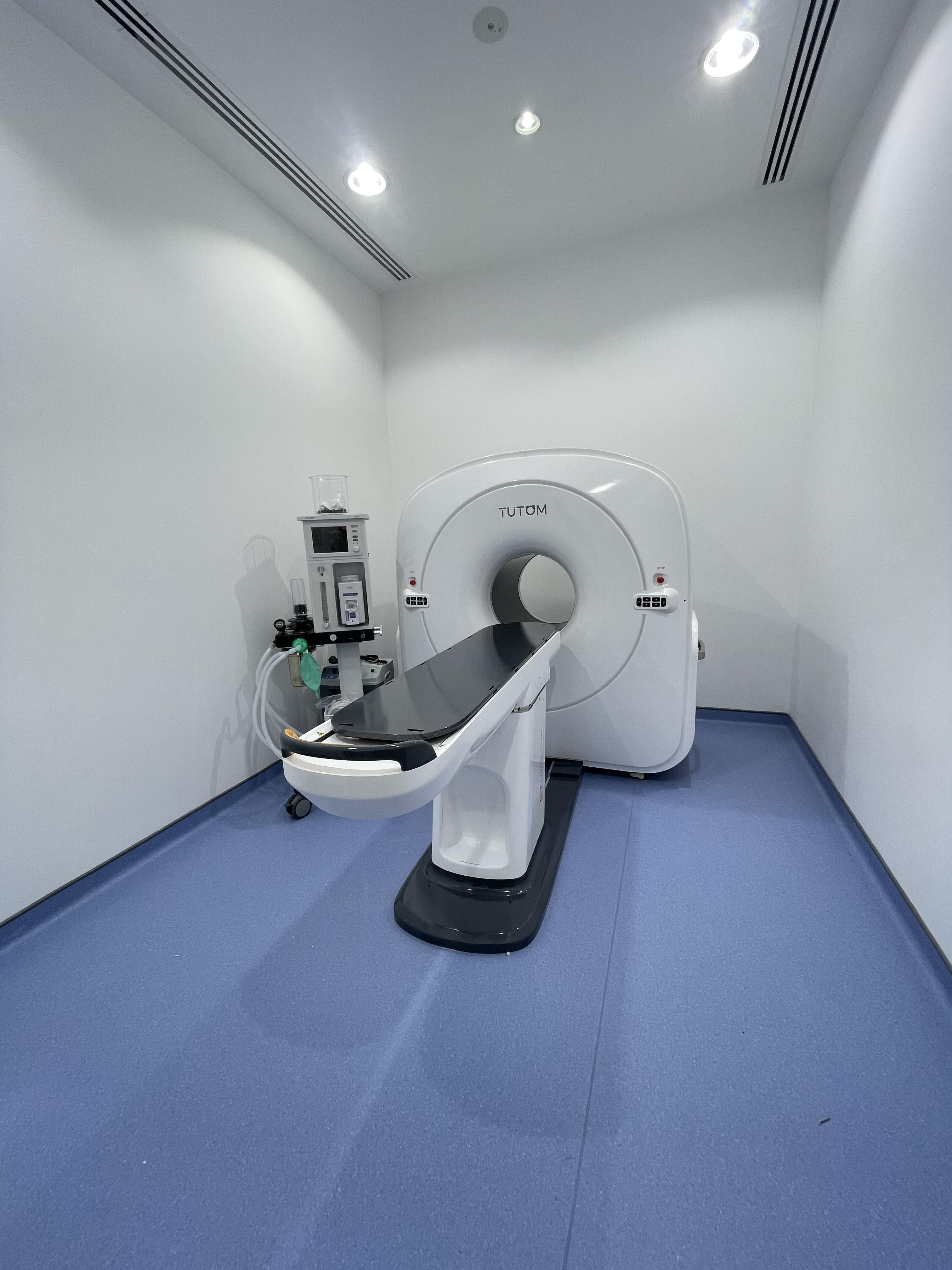
Veterinary Cone-beam CT is an innovative small-to-medium animal imaging system developed independently. It offers rapid 3D imaging through a single rotation, smart operations, advanced clinical functions and minimal radiation exposure. This cutting-edge equipment is ideally suited for capturing high-quality 3D images of bone tissue, and can also image soft tissue, making it an invaluable tool for veterinarians.
Features:
Excellent Imaging Quality:
- Amorphous silicon cesium iodide flat panel detector with 120μm pixel size.
- A slice thickness of 0.24 mm, 1.7Lp/mm high spatial resolution, and rich details.
- Small double focal spot to ensure image quality.
Smart Operation:
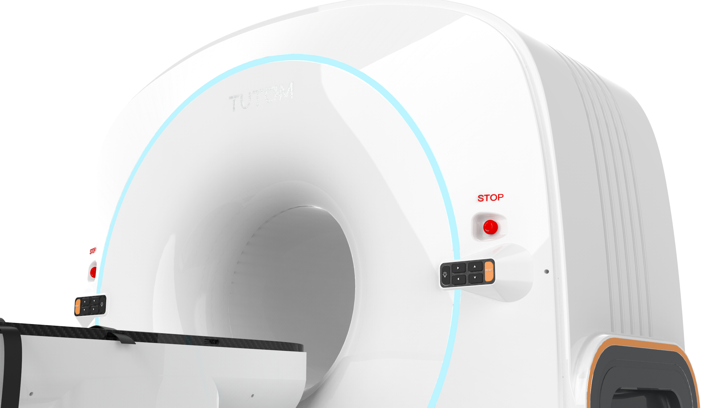
- 4 types of LED lights to indicate the machine’s status
- Laser-assisted pet positioning
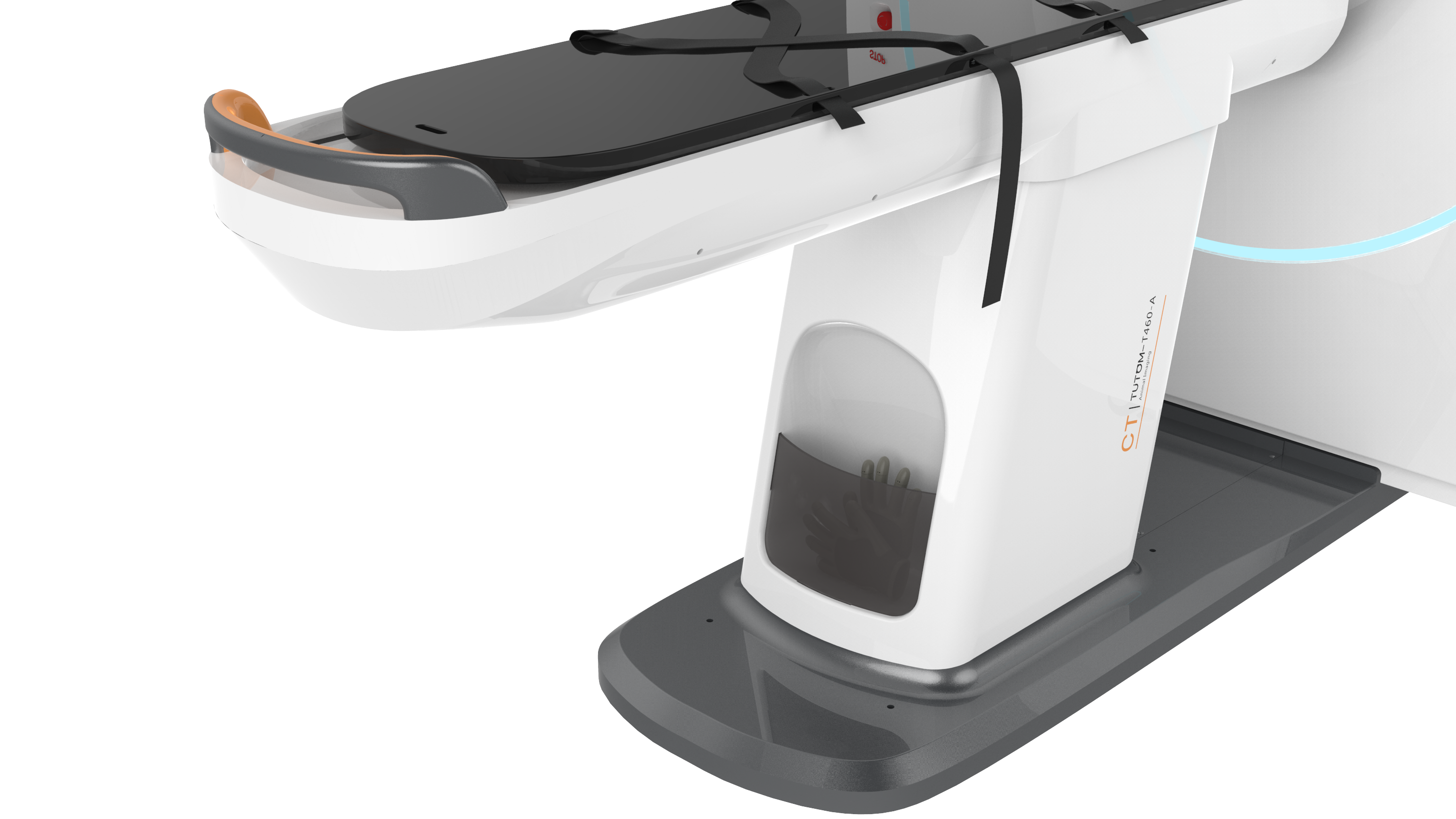
- Motorized table moving by one touch
- APR function for quick adjustment of parameter
Easy Installation:
- Compatible with 110v/220v input power
- 260kg Low weight and 10m2 small footprint
Advanced Clinical Applications:
- Radiography, contrast, and fluoroscopy all in one
- Maximum Intensity Projection
- Multiplanar Reconstruction
- Curved Planar Reformation
- VR – Skeletal Reconstruction
Technical Specifications:
| Flat Panel Detector | Scintillator | CsI |
| Image Sensor | a-Si (Amorphous Silicon) TFT | |
| Spatial Resolution | 1.7 lp/mm | |
| Pixel pitch | 120 um | |
| Slice thickness | 0.24 mm | |
| A/D | 16 bit | |
| X-ray Tube | Nominal focal spot value | Small Focus 0.3 mm Large Focus 0.6 mm |
| Tube voltage/kV | 50 kV~125 kV | |
| Tube current/mA | 2 mA~16 mA | |
| Output power | 16 kW | |
| Frame Structure | Scanning aperture | 460 mm |
| SID | 805 mm | |
| Weight | 260 kg | |
| Scanning frame size | Length, width and height: 1670 mm × 810 mm × 1600 mm, deviation±5% |
|
| Bed Structure | Bed size | Length, width and height: 1850 mm × 550 mm × 730 mm, deviation±5% |
| Maximum bearing | 60 kg | |
| Bed surface transverse range | 800 mm deviation±5% | |
| Bed lifting range | 150 mm deviation±5% | |
| Scanning parameter | 3D scanning field of view | 17 x 20 cm |
| Number of Scanning layers | 520 | |
| 2D scanning field of view | 250 x 300 mm | |
| 2D dynamic imaging frame rate | 30 fps | |
| Scanning time | 19 s |

